About

Saturday 27 July 2019

Plant Pathology Introduction

Plant Pathology Introduction 

Importance of the Plant Diseases

Globally, enormous losses of the crops are caused by the plant diseases. The loss can occur from the time of seed sowing in the field to harvesting and storage. Important historical evidences of plant disease epidemics are Irish Famine due to late blight of potato (Ireland, 1845), Bengal famine due to brown spot of rice (India, 1942) and Coffee rust (Sri Lanka, 1967). Such epidemics had left their effect on the economy of the affected countries.

Objectives of Plant Pathology

Plant Pathology (Phytopathology) deals with the cause, etiology, resulting losses and control or management of the plant diseases. The objectives of the Plant Pathology are the study on:
i. the living entities that cause diseases in plants;
ii. the non-living entities and the environmental conditions that cause disorders in plants;
iii. the mechanisms by which the disease causing agents produce diseases;
iv. the interactions between the disease causing agents and host plant in relation to overall environment; and
v. the method of preventing or management the diseases and reducing the losses/damages caused by diseases.

Scope of Plant Pathology:

Plant pathology comprises with the basic knowledge and technologies of Botany, Plant Anatomy, Plant Physiology, Mycology, Bacteriology, Virology, Nematology, Genetics, Molecular Biology, Genetic Engineering, Biochemistry, Horticulture, Tissue Culture, Soil Science, Forestry, Physics, Chemistry, Meteorology, Statistics and many other branches of applied science.

Concept of Plant Disease:

The normal physiological functions of plants are disturbed when they are affected by pathogenic living organisms or by some environmental factors. Initially plants react to the disease causing agents, particularly in the site of infection. Later, the reaction becomes more widespread and histological changes take place. Such changes are expressed as different types of symptoms of the disease which can be visualized macroscopically. As a result of the disease, plant growth in reduced, deformed or even the plant dies.
When a plant is suffering, we call it diseased, i.e. it is at ‘dis-ease’. Disease is a condition that occurs in consequence of abnormal changes in the form, physiology, integrity or behaviour of the plant. Disease is a deviation from normal functioning of physiological processes of sufficient duration or intensity to cause disturbance or cessation of vital activities.
A plant is diseased when it is continuously disturbed by some causal agent that results in abnormal physiological process that disrupts the plants normal structure, growth, function or other activities. This interference with one or more plant’s essential physiological or biochemical systems elicites characteristic pathological conditions or symptoms.

Causes of Plant Diseases

Plant diseases are caused by pathogens. Hence a pathogen is always associated with a disease. In other way, disease is a symptom caused by the invasion of a pathogen that is able to survive, perpetuate and spread. Further, the word “pathogen” can be broadly defined as any agent or factor that incites ‘pathos or disease in an organism or host. In strict sense, the causes of plant diseases are grouped under following categories:

1. Animate or biotic causes:

Pathogens of living nature are categorized into the following groups.
(i) Fungi
(ii) Bacteria
(iii) Phytoplasma
(iv) Rickettsia-like organisms
(v) Algae
(vi) Phanerogams
(vii) Protozoa
(viii) Nematodes
2. Mesobiotic causes :
These disease incitants are neither living or non-living, e.g.
(i) Viruses
(ii) Viroides

3. Inanimate or abiotic causes:

In true sense these factors cause damages (any reduction in the quality or quantity of yield or loss of revenue) to the plants rather than causing disease. The causes are:
(i) Deficiencies or excess of nutrients (e.g. ‘Khaira’ disease of rice due to Zn deficiency)
(ii) Light
(iii) Moisture
(iv) Temperature
(v) Air pollutants (e.g. black tip of mango)
(vi) Lack of oxygen (e.g. hollow and black heart of potato)
(vii) Toxicity of pesticides
(viii) Improper cultural practices
(ix) Abnormality in soil conditions (acidity, alkalinity).

Classification of Plant Disease

To facilitate the study of plant diseases they are needed to be grouped in some orderly fashion. Plant diseases can be grouped in various ways based on the symptoms or signs (rust, smut, blight etc.), nature of infection (systemic or localized), habitat of the pathogensmode of perpetuation and spread (soil-, seed- and air-borne etc.), affected parts of the host (aerial, root disease etc.), types of the plants (cereals, pulses, oilseed, ornamental, vegetable, forest diseases etc.). But the most useful classification has been made based on the type of pathogens that cause plant diseases. Since this type of classification indicates not only the cause of the disease, but also the knowledge and information that suggest the probable development and spread of disease along with their possible control measures. The classification is as follows:

1. Infectious plant diseases:

a. Disease caused by parasitic organisms: The organisms included in animate or biotic causes can incite diseases in plants.
b. Diseases caused by viruses and viroids.

2. Non-infectious or non-parasitic or physiological diseases:

The factors included in inanimate or abiotic causes can incite such diseases in plants under a set of suitable environmental conditions.

Some important terms:

1. Parasite: An organism living upon or in another living organism (the host) and obtaining the food from the invading host.
2. Pathogen: An entity, usually a micro-organism that can cause the disease.
3. Biotroph: A plant pathogenic fungus that requires living host cells i.e. an obligate parasite.
4. Hemibiotroph: A plant pathogenic fungus that initially requires living host cells but after killing the host cell grows on the dead and dying cells.
5. Necrotroph: A pathogenic fungus that kills the host and survives on the dying and dead cells.
6. Pathogenicity: The relative capability of a pathogen to cause disease.
7. Pathogenesis: It is a process caused by an infectious agent (pathogen) when it comes in contact with a susceptible host.
8. Virulence: The degree of infectivity of a given pathogen.
9. Infection: The initiation and establishment of a parasite within a host plant.
10. Primary infection: The first infection of a plant by the over wintering or over summering of the pathogen.
11. Inoculum: That portion of pathogen which is transferred to plant and cause disease.
12. Invasion: The penetration and spread of a pathogen in the host.
13. Colonization: The growth of a pathogen, particularly a fungus, in the host after infection is called colonization.
14. Inoculum potential: The growth or threshold of fungus available for colonization at substratum (host).
15. Symptoms: The external and internalreaction or alterations of a plant as a result of disease.
16. Incubation period: The period of timebetween penetration of a pathogen to the host and the first appearance of symptoms on the plant.
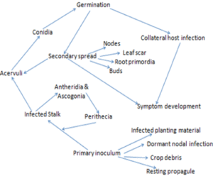
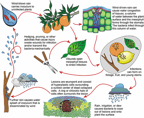
17. Disease cycle: The chain of events involved in disease development.
18. Disease syndrome: The set of varying symptoms characterizing a disease are collectively called a syndrome.
19. Single cycle disease (Monocyclic): This type of disease is referred to those caused by the pathogen (fungi) that can complete only one life cycle in one crop season of the host plant. e.g. downy mildew of rapeseed, club root of crucifers, sclerotinia blight of brinjal etc.
20. Multiple cycle disease (Polycyclic): Some pathogens specially a fungus, can complete a number of life cycles within one crop season of the host plant and the disease caused by such pathogens is called multiple cycle disease e.g. wheat rust, rice blast, late blight of potato etc.
21. Alternate host: Plants not related to the main host of parasitic fungus, where it produces its different stages to complete one cycle (heteroecious).
22. Collateral host: The wild host of same families of a pathogen is called as collateral host.
23. Predisposition: The effect of one or more environmental factors which makes a plant vulnerable to attack by a pathogen.
24. Physiologic race: One or a group of microorganisms similar in morphology but dissimilar in certain cultural, physiological or pathological characters.
25. Biotype: The smallest morphological unit within a species, the members of which are usually genetically identical.
26. Symbiosis: A mutually beneficial association of two or more different kinds of organisms.
27. Mutualism: Symbiosis of two organisms that are mutually helpful or that mutually support one another.
28. Antagonism: The counter action between organisms or groups of organisms.
29. Mutation: An abrupt appearance of a new characteristic in an individual as a result of an accidental change in genes present in chromosomes.
30. Disease: Any deviation in the general health, or physiology or function of plant or plant parts, is recognized as a disease.
31. Cop Damage: It is defined as any reduction in the quality or quantity of yield or loss of revenue resulting from crop injury.
32. Deficiency: Abnormality or disease caused by the lack or subnormal level of availability of one or more essential nutrient elements.

Effect of pathogen on the plants:

During the course of pathogenesis, normal activities of the infected host plant undergo malfunction. Consequently, morphological and physiological changes occur.

A. Morphological or structural changes:

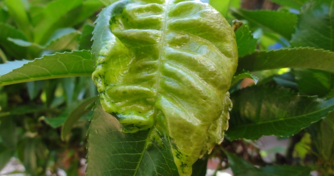
Physiological malfunctioning of the host cells causes disturbances in chemical reactionwhich ultimately lead to some structural changes viz., overgrowth, phyllody(abnormal development of floral parts into leafy structures), sterile flowers, hairy roots, witches broom, bunchy top, crown gallroot knotleaf curling, rolling, puckering etc.

B. Physiological changes:

i. Disintegration of the tissues by the enzymesof the pathogen.
ii. Effect on the growth of the host plant due to growth regulators produced by the pathogen or by the host under the influence of the pathogen.
iii. Effect on uptake and translocation of water and nutrients.
iv. Abnormality in respiration of the host tissues due to disturbed permeability of cell membrane and enzyme system associated with respiration.
v. Impairing the phenomenon of photosynthesis due to loss of chlorophyll and destruction of leaf tissue.
vi. Effect on the process of translation and transcription.
vii. Overall reproduction system of the host.

Symptoms of Plant Diseases

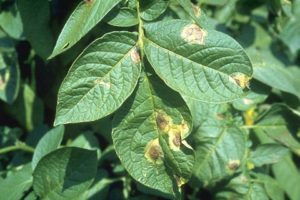
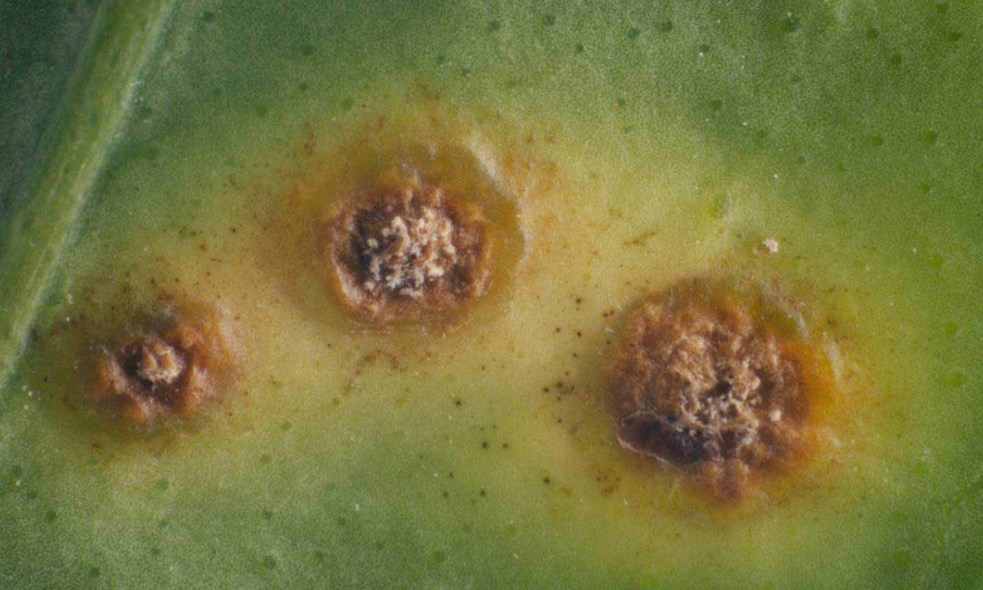
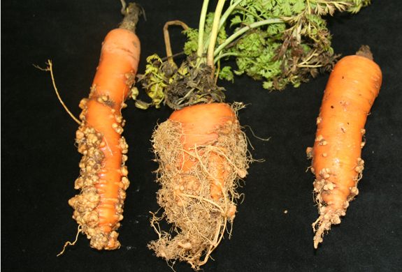
A visible or detectable abnormality expressed on the plant as a result of disease or disorder is called symptom. The totality of symptoms is collectively called as syndrome while the pathogen or its parts or products seen on the affected parts of a host plant is called sign. Different types of disease symptoms are cited below:
Necrosis: It indicates the death of cells, tissues and organs resulting from infection by pathogen. Necrotic symptoms include spots, blights, burn, canker, streaks, stripes, damping-off, rot etc.
Wilt: Withering and drooping of a plant starting from some leaves to growing tip occurs suddenly or gradually. Wilting takes place due to blockage in the translocation system caused by the pathogen.
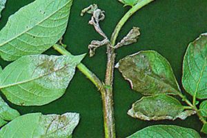
Die-back: Drying of plant organs such as stem or branches which starts from the tip and progresses gradually towards the main stem or trunk is called die-back or wither tip.
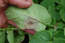
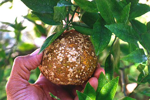
Mildew: White, grey or brown coloured superficial growth of the pathogen on the host surface is called mildew.
Rusts: Numerous small pustules growing out through host epidermis which gives rusty (rust formation on iron) appearance of the affected parts.
Smuts: Charcoal-like and black or purplish-black dust like masses developed on the affected plant parts, mostly on floral organs and inflorescens are called smut.
Blotch: A large area of discolouration of a leaf, fruit etc. giving a blotchy appearance.
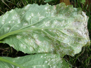
White blisters: Numerous white coloured blister-like ruptures are surfaced on the host epidermis that forms powdery masses of spores of fungi. They are called white blisters or white rust.
Colour change: It denotes conversion of green pigment of leaves into other colours mostly to yellow colour, in patches or covering the entire leaves.
(i) Etioliation: Yellowing due to lack of light,
(ii) Chlorosis: Yellowing due to infection viruses, bacteria, fungi, low temperature lack of iron etc.
(iii) Albino: Lack of any pigment and turned into white or bleached
(iv) Chromosis: Red, purple or orange pigmentation due to physiological orders etc.
Exudation: Such symptom is commonly found in bacterial diseases when masses of bacterial cells ooze out to the surface of affected plant parts and form some drops or smear, it is called exudation. This exudation forms a crust on the host surface after drying.
Overgrowth: Excessive growth of the plant parts due to infection by pathogens. Overgrowth takes place by two processes
(i) Hyperplasia: abnormal increase in size due to excessively more cell division
(ii) Hypertrophy: abnormal increase in size or shape due to excessive enlargement of the size of cell of a particular tissue.
Atrophy: It is known as hypoplasia or dwarfing which is resulted from the inhibition of growth due to reduction in cell division or cell size.
Sclerotia: These are dark and hard structures of various shaped composed of dormant mycelia of some fungi. Sometimes, sclerotia are developed on the affected parts of the plant. Presence of sclerotia on the host surface is specifically called a sign of disease rather than symptom.
Development of epidemics:Sudden outbreak of a disease within a relatively short period covering a large areaand affecting many individuals in a population is called epidemic. Although, this term was originally designated to the human diseases, now applied in the diseases of animals, poultry, plants etc. Epidemic form of plant diseases is called as epiphytotics.
For a disease to occur, coincidence of three parameters of disease triangle is essential, namely, the vulnerable hostvirulent pathogenand favourable environment. Under such circumstances, the pathogen not only completes its life cycle but also undergoes repeated generations. Then an epidemic develops only when few repeated generations are completed by the pathogen on the same host. As each generation or cycle of the pathogen takes a few days for completion, the fourth parameter i.e. time factor (forms a disease tetrahedron or disease pyramid) is also involved in epidemic build up.
In other words, epidemic growth is both temporal (pertaining to time) and a spatial(relating to space or area) process. The initial stages of an epidemic growth curve have a lag phase, when the incubation period is longer, inoculum load is weak and prevalent environmental conditions are unfavourable. Subsequently, when the conducive conditions occur, the growth of the disease is rapid and the severity of the epidemic explodes like a time bomb. Later, severity declines either due to unfavourable weather or crop maturity or both.
Plant Disease Management:
The word ‘control’ is a complete term where permanent ‘control’ of a disease is rarely achieved whereas, ‘management’ of a disease is a continuous process and is more practical in influencing adverse affect caused by a disease. Disease management requires a detail understanding of all aspects of crop production, economics, environmental, cultural, genetics and epidemiological information upon which the management decisions are made.

A. Principles of plant disease management:

There is six basic concept or principles or objectives lying under plant disease management.
1. Avoidance of the pathogen: Occurrence of a disease can be avoided by planting/sowing a crop at times when, or in areas where, inoculum remain ineffective/inactive due to environmental conditions, or is rare or absent.
2. Exclusion of the pathogen: This can be achieved by preventing the inoculum from entering or establishing in a field or area when it does not exist. Legislative measures like quarantine regulations are needed to be strictly applied to prevent spread of a disease.
3. Eradication of the pathogen: It includes reducing, inactivating, eliminating or destroying inoculum at the source, either form a region or from an individual plant (rouging) in which it is already established.
4. Protection of the host: Host plants can be protected by creating a toxin barrier on the host surface by the application of chemicals.
5. Disease resistance: Preventing infection or reducing the effect of infection of the pathogen through the use of resistance host which is developed by genetic manipulation or by chemotherapy.
6. Therapy: Reducing severity of a disease in an infected individual. The first five principles are prophylactic (preventive) procedure and the last one is curative.

B. Methods of plant disease management

1. Avoidance of the pathogen:
i. Choice of geographical area
ii. Selection of a field
iii. Adjustment of time of sowing
iv. Use of disease escaping varieties
v. Use of pathogen-free seed and planting material
vi. Modification of cultural practices

2. Exclusion of inoculum of the pathogen

i. Treatment of seed and plating materials
ii. Inspection and certification
iii. Quarantine regulations
iv. Eradication of insect vector

3. Eradication of the pathogen

i. Biological control of plant pathogens
ii. Eradication of alternate and collateral hosts
iii. Cultural methods:
a. Crop rotation
b. Sanitation of field by destroying/burning crop debris
c. Removal and destruction of diseased plants or plant parts
d. Rouging iv. Heat and chemical treatment of diseased plants
v. Soil treatment: by use of chemicals, heat energy, flooding and fallowing

4. Protection of the host

i. Chemical control: application of chemicals (fungicides, antibiotics) by seed treatment, dusting and spraying
ii. Chemical control of insect vectors
iii. Modifications of environment
iv. Modification of host nutrition

5. Disease resistance Use of resistant varieties:

Development of resistance in host is done by
i. Selection and hybridization for disease resistance
ii. Chemotherapy
iii. Host nutrition
iv. Genetic engineering, tissue culture

6. Therapy Therapy of diseased plants can be done by

i. Chemotherapy
ii. Heat therapy
iii. Tree-surgery

White Rust of Crucifers| Albugo| Disease cycle| Control Measures|

White Rust of Crucifers| Albugo| Disease cycle| Control Measures|

HOSTS

White rust most commonly occurs on: field mustard (Brassica campestris L.), leaf or Chinese mustard (B. juncea Zerj, & Coss.), black mustard (B. nigra (L.) Koch), broccoli and cauliflower (B. oleracea L. var. botrytis L.), Chinese or celery cabbage (B. pekinensis (Lour.) Rupr.), rutabaga (B. campestris L. var. napobrassica (L.) DC.), pak-choi (B. chinensis L.), turnip (B. rapa L.), radish (Raphanus sativus L.), and daikon (R. sativus L. var. longipinnatus Bailey). Less common hosts are spinach (Spinacia oleracea L.), horseradish (Armoracia lapthifolia Gilib.), and “pepper grass” (Lepidium L. sp.).
white-rust-of-crucifers
DISTRIBUTION
  1. candida(syn.:A. cruciferum) occurs in all parts of the world where cruciferous crops are grown.

SYMPTOMS

White rust is a common but usually not serious disease. It is characterized by both local and systemic symptom expression. Local infection appears as pustules or “blisters” filled with sporangia on leaves, smaller stems, and floral parts. Pustules measure approximately 1 to 2 mm in diameter and are white or creamy yellow. Systemic infections result in abnormal growth, distortion, and sterility of flowers or inflorescences. These abnormalities (or hypertrophy) are known as stagheads and consist mostly of thick-walled oospores.

BIOLOGY

Races:
To date, seven distinct A. candida races are known based on susceptibility to 7 different host species, with each species susceptible to its own specific race. Young seedlings, without fully expanded cotyledons, usually are susceptible to all races while older plants usually are race specific. Most of the Albugo candida races have a broad host range. In a study in which 115 varieties of 10 Brassica species were inoculated with A. candida race 1 (from Raphanus sativus) and 2 (isolated from B. juncea), all varieties but one showed some degree of susceptibility to one or both races. Cultivars within species varied in susceptibility to both pathogenic races.

Sporangial Germination:

Sporangia are produced in pustules and once liberated, are dispersed by wind, rain, or insectsto neighboring plants. The sporangia require some drying in order to germinate well. Each germinating sporangium gives rise to five to seven zoospores. The preferred temperature for germination ranges from 1 to 18 ºC, but is optimum between 10 ºC and 14 ºC. Temperatures should be between 16 ºC and 25 ºC, with the optimum at 20 ºC for zoospores to produce germ tubes and penetrate plant tissue. The moisture necessary for zoospore activity is ideal when in the form of heavy dew or fog or during periods of extended rainfall and lower temperatures.
When systemic infection of inflorescences occurs, stagheads containing numerous thick-walled oospores are produced resulting in serious losses in seed yield. Formation of stagheads is favored when the environmental conditions include little sunlight (2-6 hours per day) and extended rainfall (up to 161 mm) during the flowering period.
Oospore germination occurs in one of three ways:
1) production of a sessile vesicle,
2) an exit tube with a terminal vesicle,
3) or a germ tube.
The most common type of germination (approximately 70% of the time) is the production of a sessile vesicle, which releases 40 to 60 zoospores. Germination can be increased in vitro by “washing” oospores with water in flasks on a rotary shaker. This method is consistent with the general observation that germination is enhanced by the leaching action in spring water from melting snow or rain showers in more temperate climates.
There is some evidence of heterothallism in A. candida. One study examined the pathogen on a host species and noticed that primary infections were sterile but became fertile when secondary infections were established. The hypothesis that isolates of Albugo are self-sterile, but fertile when they interact with a compatible isolate, was supported by (the) observation that oospore production in Albugo is frequently correlated with a secondary infection by the related pathogen, Peronospora parasitica. It is believed that Peronospora parasitica stimulates gametangium formation in Albugo, and subsequent oospore formation by selfing.
Infection:
Oospore contaminated seeds and zoospore infected flower buds perhaps are the most important sources of primary infection of white rust. In the laboratory, inoculation of cotyledons and leaves produce few stagheads, while inoculations of flower buds result in approximately 55% infection. In the field, flower buds may be infected in one of two ways, by infested seeds where the pathogen develops systemically or by infection by sporangia from other plants. Oospores on seeds are able to survive for a long period of time when kept dry, but do not survive as long in the soil. In one study oospores could not be recovered from white rust infested fields six months after harvest. This study examined infected plant debris in the soil and buried hypertrophied host tissue, suggesting that oospores in field soils are not a primary source of infection for the next crop.
albugo-life-cycle

EPIDEMIOLOGY

White rust is spread through oospore contaminated seeds, wind and rain borne zoosporangia, and possibly perennial mycelium in infected plants. Non-cruciferous weeds are not believed to serve as sources of inoculum.

MANAGEMENT

NON-CHEMICAL CONTROL

Plowing or disking diseased plants and plant parts results in rapid decomposition of infected tissues and helps to significantly reduce future white rust infection. Crop rotation with noncruciferous host plants is also effective. Weed control and other sanitary methods are necessary too.

Resistance:

Resistance has been successfully deployed with mustard and rutabaga, however, with Asian vegetables such as Chinese mustard, Chinese cabbage, pak choi, and diakon, resistant varieties have not yet been identified.

CHEMICAL CONTROL

The development of the acylalanine fungicide metalaxyl (Ridomil; Subdue) greatly improved the ability to control while rust with fungicide application. Metalaxyl provides limited curative activity and some control of systemic infection.
Applications should be made to the soil and subsequently applied to the foliage. Frequency of application would vary according to the length of crop and amount of rainfall experienced. In temperate environments a soil application and a minimum of 1-2 foliar applications during the crop cycle is suggested.
With the possibility of developing fungicide tolerant pathogen strains associated with metalaxyl, growers should consider using Ridomil MZ58 formulations with foliar fungicide applications. This formulation adds a second fungicide to the tank mix.
Older fungicides used, but less effective, for white rust control include: Dithane Z-78, Blitox, wettable sulphur, fixed copper compounds, Bordeaux mixture, chlorothalonil, captofol, captan, dodine, mancozeb, metiram, maneb, and zineb.

Rust of Wheat

Rust of Wheat

Introduction:
Rust diseases of wheat are among the oldest plant diseases known to humans. Early literature on wheat cultivation mentions these devastating diseases and their ability to destroy entire wheat crops. Since rust discovery, numerous studies have been conducted on the life cycles of rust pathogens and their management. The information gained from these studies has enabled us to develop best management practices that reduce the impact of the diseases. Today, worldwide epidemic losses are rare, though the diseases can occur at significant levels in particular fields or throughout a particular growing region. The persistence of rust as a significant disease in wheat can be attributed to specific characteristics of the rust fungi. These characteristics include a capacity to produce a large number of spores—which can be wind-disseminated over long distances and infect wheat under favorable environmental conditions—and the ability to change genetically, thereby producing new races with increased aggressiveness on resistant wheat cultivars.
Symptoms:stem-rust-exampleThere are three rust diseases that occur on wheat: stem rust, leaf rust and stripe rust. These diseases are each caused by a particular species of the “rust” fungus, Puccinia. Rust fungi all produce similar disease symptoms on the host plants and have similar requirements for infection. The diseases get their name from their appearance on the plant. Infection can occur on any above-ground plant part, leading to the production of pustules that contain thousands of dry yellow-orange to reddish-brown or black spores. These pustules give the appearance of “rust” on the plant.
Stem rust occurs primarily on stems but can also be found on leaves, sheaths, glumes, awns, and even seed.glumes-rust
Symptoms begin as oval to elongate lesions that are generally reddish-brownin color. In the late stages of the disease, erumpent pustules produce numerousblack sooty spores. Severe infestations with many stem lesions may weaken plant stems and result in lodging.
Symptoms begin as small circular to oval yellow spots on infected tissue of the upper leaf surface. As the disease progresses, the spots develop into orange-colored pustules that may be surrounded by a yellow halo.
The pustules produce a large number of spores that are easily dislodged from the pustule, resulting in an “orange dust” on the leaf surface or on clothes, hands, and equipment. As the disease progresses, black spores may be produced, resulting in a mixture of orange and black lesions on the same leaf. Tiny orange lesions may be present on seed heads, but these lesions do not develop into erumpent pustules. This difference helps to distinguish leaf rust from stem rust.rust-leaves-chlorosis
Chlorosis, or yellowing, of leaves can be quite evident with both leaf and stripe rust, and fields with plants displaying severe symptoms may be easily detectable from a distance.
Causal Organisms:Stem rust (also known as black stem rust) is caused by Puccinia graminis f. sp. tritici.It is primarily a disease on wheat, though it can also cause minor infections on certain cultivars of barley and rye. Leaf rust is caused by Puccinia recondita f. sp. tritici(now known as Puccinia triticina). Like the stem rust fungus, this pathogen is primarily a problem on wheat, but it may be weakly pathogenic on some cultivars of barley, triticale, and some species of goatgrass and wheatgrass.
Life Cycle:
Rust fungi have complex life cycles, which may require two specifically different host plants and up to five different spore stages. Rust diseases that require two host plants to complete the life cycle generally have what is known as an economic host and an alternate host. The economic host, in these diseases, is wheat. The alternate host is typically a weed or native plant . For example, barberry (Berberis vulgaris) is the primary alternate host for the stem rust fungus. Infection of barberry results in circular, yellow- to red-colored pustules on the underside of the leaves. Spores (aeciospores) produced on barberry plants infect wheat, and another type of spore (basidiospores) produced on wheat infects barberry plants. Although both hosts are necessary to complete the full life cycle, epidemics on wheat can develop rapidly because spores (uredospores) produced on wheat can cause auto-infection (spores infect the same plants on which they were produced). This spore stage of the life cycle is known as the repeating stage and is responsible for the rapid development of disease outbreaks.
life-cycle-of-rust
Aecia on the surface of barberry
Disease Development:
Water on the leaf surface from intermittent rains or heavy dews and temperatures conducive for germination and growth of the pathogen are required for disease development. Stem rust is a warm-temperature disease that develops optimally between 65 and 85°F; however, the disease can occur at temperatures between 59 and 104°F. Leaf rust develops optimally at temperatures between 59 and 71°F, and the disease will progress until temperatures are above 80°F. Stripe rust is a lower-temperature disease that is generally found at higher elevations and in cooler climates. The optimal temperature for the development of this disease is 50 to 59°F, with disease progression ceasing at temperatures above 70°F. When conditions are optimal for disease development, infection is completed in 6–8 hours and uredospores capable of causing secondary spread of the disease are produced in 7–10 days. Uredospores are relatively limited in the length of time they remain viable compared to other spore stages produced by rust fungi. Yet they are extremely efficient in spreading disease because they are produced in large quantities and are easily spread by wind. In locations with mild winters, uredospores have been found to survive year-round. Therefore, alternate hosts and some spore stages are required to complete the pathogen’s life cycle but are not needed for the initiation of new infections each year.
Prevention:
One of the only early prevention methods for avoiding rust diseases or minimizing their impact is to plant a variety with known resistance. Variety resistance is the most economical method of control. In some cases, avoiding rust is not possible because of constant changes in strains (races) of the pathogens.
Eradication of barberry plant is an important mean to eliminate the disease.
Application of fungicides is very convenient method to control rust.

Loose smut of wheat| Causal Organism| Ustilago

Loose smut of wheat| Causal Organism| Ustilago
It is one of the common disease of wheat caused by Ustilago tritici.  Smut fungi belonging to order Ustilaginales are parasites both on monocots and dicots. Since they impart sooty appearance due to production of black spore mass they are generally described as smut fungi.
The mycelium of these fungi is parasitic dikaryotic and quite often intercellular in the host plant, may be systemic or localized. Ustilaginales as a group do not possess orgainsed sex- organs but dikaryotization is brought about by the fusion of mycelial hyphae or by fusion of basidiospores or sporidia. Chlamydospores, the reproductive structures have been designated variably

Symptoms:

The affected plant is converted into a black mass of spores and no grains are formed. The infection brings about physiological and structural changes in the host.
In most of the varieties under cultivation the growth of the plant and its general appearance is not affected in any way until ears have appeared. But in some varieties, abnormalities are seen in the flag leaves.
The disease is called loose smut because the spores produced on smutted heads are easily removed by wind and rain. Typically, after a short while, only the bare, sooty, flower stocks remain. In some cases, the seed head is only partially infected. Prior to heading, diseased plants may have dark green leaves with chlorotic streaks, but this symptom generally goes unnoticed. Seed with no visible symptoms can be infected with the fungus.

Disease Cycle:

The fungus survives as dormant mycelium in symptomless seed. When infected seed germinates, the fungus systemically infects the plant. Seed produced on infected plants are converted to fruiting bodies containing copious amounts of spores.Wind, rain and insects disseminate the spores. to neighboring plants. Plants are susceptible to infection only for one week during flowering. Open flowers become infected when spores germinate and penetrate the ovary wall. Seed that develops from infected ovaries appear normal but harbor the pathogen. These infected seed are fully germinable and serve as primary inoculum for new disease outbreaks if used for planting.

Conditions for Disease:

Infection occurs only during flowering and is favored by humid conditions with cool to moderate temperatures (60 – 72 °F). These conditions coupled with light showers or heavy dews are particularly favorable for disease development.

Management:

Before the advent of systemic fungicides, hot water or solar energy methods of seed treatment were the only recommendations. The heat therapy is effective in the case of internally seed-borne diseases where the mycelium remains in the embryo in the dormant form. Hot water treatment of seed at 49ºC for 2 min eliminates mycelium of the fungus from infected embryos.
Seed is soaked in ordinary water for 4 h (8 a.m. to 12 noon) on a bright summer day (May-June) and spread in a thin layer and dried in the sun for 4 h (12 noon to 4 pm.)
The disease is primarily managed by the use of resistant cultivars and pathogen-free seed. Although many cultivars exist with resistance to some races of the fungus, no cultivars are resistant to all races. Therefore, cultivar resistance alone isn’t sufficient to manage the disease.
Seed production fields must be inspected at heading to ensure the absence of loose smut.
Systemic fungicides can also be used as a seed treatment to help control the disease in germinating seedlings. No treatments are available to protect flowers from wind-blown or rain-splashed spores.

Red Rot of Sugarcane -

Red Rot of Sugarcane 

Introduction:

Red rot, caused by Colletotrichum falcatum, is one of the major diseases of sugarcane in the world.

Symptoms:

Red rot attacks the stalks, stubble rhizomes, and leaf midribs of thesugarcane-glom
sugarcane plant. It may invade leaf-blade and leaf-sheath tissues
and is capable of infecting sugarcane roots but it is not
important as a disease of these organs.
Red rot is frequently not discernible from external examination of
the sugarcane stalk unless it has so completely rotted the interior as to
cause the rind to lose its natural bright color and become dull in
appearance. Plants so affected may be detected by the yellowing, shriveling, and dying of the upper leaves. The disease is
recognized by the longitudinal reddening of the normally white or
yellowish-white internal tissues2192_0of the internode, especially when this red color is interrupted by occasional white patches extending crosswise of the stalk.

Life cycle and disease cycle:

Manifestation of red rot varies depending on the nature of infection, time of the season and the prevailing environment. If sufficient inoculum is present in/on the set in active form and if adequate moisture is available, it causes both pre and post-emergence mortality of the sprouts during April-May in the subtropical region. With the advent of pre-monsoonshowers, symptoms of the disease start appearing, and with the onset of the monsoon when the weather is most suitable, full manifestation of the disease takes place. Later on, infected plants turn yellow and finally dry up. At the grand growth phase, when sufficient stalk and sugar have formed, typical stalk rot phase (red rot phase) appears. Red rot is not easily identified from the external appearance of the cane unless it has caused irreparable damage by rotting the internal tissues. In such cases, the rind loses its natural bright colour, turns violet or displays dark reddish tinge, and becomes dull in appearance. The affected plants appear sick; usually yellowing of the 3rd or 4th leaf, these being the prominent leaves of the crown, draw the attention of the observer from a distance (The yellowing may start from any leaf of the crown there is no hard and fast rule). Yellowing of leaf starts from the tip and proceeds down along the leaf margin. C. falcatum proliferates within the cane stalk happily, and usually comes out through the root primordia for secondary spread when sufficient rotting of the internal tissues has taken place. The fungus totally transforms the root primordia into black acervuli bearing abundant conidia (the black colour is due to the dark setae). At this stage, if the cane is split open longitudinally, typical symptoms of red rot, viz., reddening of the internal tissues with interrupted red and white patches (white spots) in the affected internodes, rotten or damaged node, and the presence of typical sourly alcoholic smell may.

Control Measures:

Adopting one or more of the following measures can minimize the disease incidence
Avoid planting of highly susceptible varieties.
Follow the long furrow method or pair row method of layout for planting and irrigation.
Planting material should be collected from the seed nursery
Before primary and general cane planting set treatment with 0-1% carbendazim (Bavistin) for 15-20 minutes dipping should be followed
To control the secondary spread of the disease, follow the following practices
Rogue out the affected clumps & destroy it by burning. Stools should not buried in the soil or should not be kept or thrown on bunds
After roguing, drench the spot with the 0.1% carbendazim containing fungicide
Foliar application of the Bavistin (0.1%), or Baynlate 70% w.p. (0.1% to 0.15%) by 2 to 3 times at an interval of 10 to 12 days may be tried. Spraying may be done immediately after the disease incidence
After the harvest of the diseased crop, left over trash with stools should be burnt immediately
Crop rotation should be followed to break the rapid built up of the disease. In rotations, it is advisable to include green manure crop. Because this not only increases the population of the antagonistic mycoflora but also helps to break the life cycle of disease
Avoid ratooning of the diseased crop
idm-of-red-rot

red-rot-of

Late Blight of Potato| Phytophthora infestans| Symptoms & Control

Late Blight of Potato| Phytophthora infestans| Symptoms & Control
Late blight is not only the most serious fungal disease of potatoes, it also occurs almost everywhere where potatoes are grown and is especially important in the traditional potato growing areas. On several occasions the disease has reached disastrous proportions. Well documented is the Irish Famine of around 1845 to 1850 when, as a result of a late blight epidemic, one million persons of a total of eight million inhabitants died and another 1.5 million left the country. The population of Ireland had completely depended on the potato that virtually formed its only food source. The biology of the disease and methods of its control were as yet entirely unknown. In other parts of Europe and in North America, the disease was as severe as in Ireland. In these areas, however, 3 famine was avoided due to a more varied food supply.
In spite of increased knowledge on the disease, late blight continues to be a major constraint to potato production worldwide. If not controlled, losses may reach 100 percent, and even lower infection levels may make the crop unfit for storage.

SYMPTOMS:

In an advanced stage of disease development, symptoms resemble those caused by frost attack. Plants severely affected with late blight produce a distinctive odor, which results from the breakdown of plant tissue. The disease affects leaves, stems, and tubers.

Leaves.

The earliest symptoms of the disease are often present on lower leaves. They consist of small, pale to dark green spots the, change into brown or black lesions, depending on the humidity of the air. Lesions begin frequently at leaf tips and margins. A pale green or yellow border, a few millimeters wide, often separates dead from healthy tissue.
Under conditions of high humidity and cool temperatures, lesions expand rapidly. Sporulation may be visible at the lower surface of the leaves as a white mildew surrounding the lesions. it may become inconspicuous during the day, when the lesions dry and shrivel. In less than a week, the disease may spread from the first infected leaflets on a few plants to most plants in a field. Leaves may drop off.

lateblight03

download

Stems:

Either by direct infection or extending from the leaves, lesions may develop on petioles and stems, where they expandlateblight04
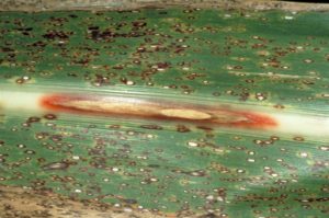
lengthwise. Infected stems are weakened, and may collapse thereby causing death of plant parts above the lesion.
Tubers:
x-late-blight
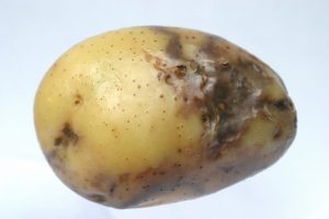
Infected tubers show a superficial and irregular discoloration. Dry and brown necrotic lesions penetrate from the surface into the tuber tissue. Secondary pathogens (mainly bacteria) may convert the almost odorless(faint vinegar smell) dry rot typical for P. infestans into a badly smelling soft rot. Late blight normally does not spread during storage, but secondary infections may contaminate other tubers.

Life Cycle of P. infestans

Most stages of the life cycle of P. infestans can only be seen with the aid of a microscope. In this way, fungal threads (mycelium) can easily be observed. They are characterized by the absence of cross walls (septa). The mycelium develops between cells (intercellularly) and only extensions of it (haustoria) enter the cells. Both asexual (vegetative) and sexual (generative) reproduction occurs.

Asexual reproduction.

Between three and ten days after infection, depending on environmental conditions, spore-bearing organs (sporangiophores)emerge through openings (stomata) on the leaf surface. Stomata are more frequent on the underside than on the upperside of leaves. This explains why sporulation on the underside is more abundant than on the upperside. Zoosporangia (also called sporangia or conidia) develop at the end of these sporangiophores. When mature, the zoosporangia easily break off and are spread by the wind. Most spores are deposited after a few meters, however, travel distances of over 30 km have been reported.potato_late_blight_life_cycle
lateblightcycle2
Sporulation can be demonstrated in a simple experiment: A stem with small lesions on the leaves is placed in a flask with water. Stem and flask are covered with a plastic bag. It is kept overnight at a temperature between 15 and 22 °C (room temperature). Next morning the white sporulation, especially on the underside of the leaves, can be observed.
The size of zoosporangia is just below the detection limit of the unaided eye. Their lemon-shaped form can he observed with a microscope. Zoosporangia may germinate directly or indirectly.
To a small degree, zoosporangia germinate directly at temperatures above 20ºC (optimum 24 °C). Zoosporangia behave as single spores. They form germ tubes that penetrate into the plant tissue. In nature, direct germination seems of little importance.
Zoosporangia germinate indirectly at temperatures of 12 to 16phytophthora-spores
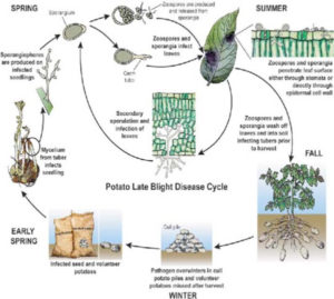
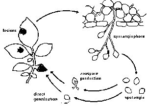
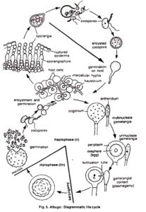
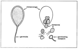
°C,each sporangium releasing from 10 to 20 swarm spores (zoospores). Activated by two flagellae, zoospores remain motile from a few minutes up to several hours. Under certain conditions they lose the flagellae, form a cell wall and subsequently a germ tube.
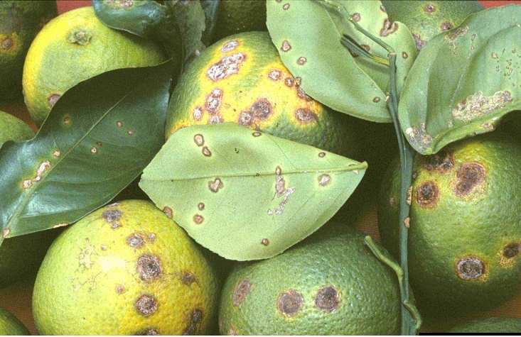
On leaves and stems, germ-tubes may directly penetrate the plant epidermis (no stomata are required). On tubers, germtubes penetrate through lenticels or wounds. Since the fungus cannot survive for a prolonged period outside the host tissue zoosporangia or zoospores die, if no suitable host tissue is available.

Sexual reproduction:

Until 1984, the sexual stage of P. infestans has only been reported in Mexico and parts of Central America. More recent reports inform about its occurrence in other parts of the world. When mycelia of different types of the fungus (called mating types A1 and A2) grow together, one of them may form male cells (antheridia) and the other female cell! (oogonia). The oogonium grows through the antheridium, allowing fertilization. The fertilized oogonium develops into a thick-walled resting spore (oospore). Oospores, in contrast to zoosporangia and zoospores, can resist unfavorable conditions, such as droughts and low temperatures.
Formation of oospores helps P. infestans and related species to survive adverse conditions, such as the winter, dry periods, and absence of hosts. Possibly due to the absence of the  mating type, oospore formation in P. infestanshas not been observed outside Mexico and Central America.
Oospores of P. infestans germinate through formation of sporangia, similar to those described in asexual reproduction. After infection of a host, the resulting zoospores may start a new life cycle.

EPIDEMIOLOGY :

With the exception of oospores which may survive in the soil, in nature the pathogen persists only in susceptible hosts. Sources of infection are:” infected seed tubers, ” cull piles, ” neighboring potato fields, ” other host plants.
Infected seed tubers. In areas where potatoes are grown during defined seasons, diseased seed tubers are often the most important source of infection. Tubers become infected through lenticel and wounds when spores are washed into the soil by rain from infected leaves, especially when tubers are formed superficially or are not well covered by hilling. Also at harvest, tubers can become contaminated by contact with infected foliage.
Blight-infected tubers normally rot when planted in the field. However, few diseased tubers may form sprouts which then become primary sources of infection.
Cull piles. Infected tubers are frequently found in cull piles. Also tubers from previous crops that have been left in the field may be infected and may form ‘ source of primary infection for a new crop.
Neighboring potato fields. Neighboring potato fields are another source of infection especially in areas where potatoes are grown during the whole year.
Other host plants. Some other solanaceousplants may become affected by P. infestans. In many countries, the ornato is the most important alternative host. The pathogen also persists on wild species of potatoes that are widely distributed in the highlands of Mexico, Central and South America.
From primary infection sources, the wind disseminates the spores towards the fields. For development of the disease, temperature and humidity are of fundamental importance.
The most favorable temperature for development of the fungus (mycelium, sporangia) is around 21 °C, but the fungus remains alive in host tissues at air temperatures between 0 and at least 28 °C. Zoosporangia develop at temperatures between 9 and 22 °C.
Development of the fungus within the leaf is little affected by air humidity. However, sporangia are only formed when the relative humidity in the leaf canopy is above 95 percent. When the temperature is optimum, about eight hours of high hunlidity are required for production of zoosporangia, liberation of zoospores, and penetration. Water (dew, rain) must be on the leaf surface for at least two hours to allow zoospore formation, germination, and penetration.
In summary, the diease develops and spreadsmost rapidly at low temperatures and high humidity. Under these conditions, a disease cycle takes only three days. Unfavorable conditions may delay or temporarily interrupt the disease cycle.
For better disease management and the planning of a spray program, several methods of late blight forecasting have been developed, and are based on observation of temperature and humidity conditions. A severe late blight attack can be expected when the “leaf wetness period” exceeds 8 to 10 hours for several consecutive days, and temperatures range between 10 and 24 °C. Typical blight weather is characterized by cool, ,loudy days with frequent rains.
Disease forecasting programs appear to function generally better in temperate climates, than in most of the tropical highlands where potatoes are mainly grown in the rainy season.

CONTROL :

Development of late blight is favored when the potato variety is susceptible, and appropriate environmental conditions persist for sufficient time. Generally, any agronomic measure that affects this relationship may be utilized to reduce the disease.
Healthy seed. A basic condition for adequate crop production is the use of non-infected seed. This eliminates the primary infection source from the field.
Planting procedure. In regions with defined rainy seasons a change in planting time may help to reduce disease severity. This might however reduce the yield, as the potato requires abundant water during tuber formation.
Crop care. Any treatment that promotes rapid drying of foliage and reduces humidity within the crop helps to restrict disease development. These include wider planting distance and appropriate irrigation procedures. Sprinkler irrigation tends to increase disease severity.
Exposed tubers and those poorly covered by soil become readily infected by fungus spores washed down from the foliage.
Proper hilling reduces the amount of spores reaching the tubers and may result in faster drying of the field after a rain.
Resistance. Varieties with the highest resistance to late blight should be used, if commercially acceptable. Two types of resistance, race specific resistance and general resistance.
Harvest. When the foliage has been affected by late blight, it should be destroyed mechanically or Chemically at least one week before harvest. This practice reduces the chance of tuber infection by contact with infected leaves and stems and promotes skin suberization, making tubers less vulnerable to infection. In addition, this helps to reduce mechanical damage and infection by storage pathogens.
Tubers should be only harvested when they are mature (the skin should not be peeling). The soil should be dry to present infection through damaged skin or lenticels. Only disease-free tubers should be stored.
Crop residues, including infected tubers, should be removed from the field or plowed under. Cull piles should be covered with sufficient soil to prevent emergence of discarded tubers.
Fungicide applications. Preventive fungicides principally inhibit spore germination and penetration. Once the pathogen enters the leaves, most fungicides are ineffective. A preventive fungicide application program should start immediately after the first blight symptoms appear in a crop. Proper fungicide cover on the foliage should be maintained as long as favorable late blight conditions exist. In a susceptible crop, up to 15 applications per season may be required.
Proper dosage and application are as important selecting an effective fungicide.
Instructions given with the fungicide should be followed carefully. The spraying equipment should be kept in good condition to Lssure an even and fine spray application.
Observation of meteorological data may help determine when fungicide applications are necessary.
Newly developed systemic fungicides (acylalanines) are translocated within the plant. A constant and uniform coverage is less critical than with conventional fungicides, and frequency of spraying may be reduced. Granular formulations applied to the soil do not require spraying equipment. Systemic fungicides may be highly effective; however, development of resistance of the fungus to these fungicides has been reported recently.
No general recommendations for fungicide application can be given, since availability, effectivity, conditions, and regulations vary from country to country. Local experts should be consulted for recommendations in a particular area.
Integrated control. The best control is a combination of preventive measures, based on the use of resistant varieties. Costly fungicide applications can then be reduced. The objective of integrated management of late blight is not the eradication of the disease, but the most economical production of a potato crop.

Monday 15 July 2019

Citrus Canker


Citrus Canker


Citrus fruits showing canker
Fig: Citrus canker
Citrus canker is one of the most feared of citrus diseases, affecting all types of important citrus crops. The disease causes extensive damage to citrus and severity of this infection varies with different species and varieties and the prevailing climatic conditions.
Citrus canker presently occurs in over thirty countries in Asia, the Pacific and Indian Ocean islands, South America, and the South eastern USA. When citrus infection occurs in the early growing stage, the fruits crack or become malformed as they grow, and the heavily infected ones fall prematurely. Light infection in later growth stages may cause only scattered canker lesions on the surface of fruits but makes fresh fruits unacceptable for market. The severity of fruit infection usually parallels that of foliage infection. Eighty to ninety percent of fruit infection is not uncommon in susceptible citrus trees that have already sustained severe foliage infection. Such heavy foliage infection often causes severe defoliation, leaving only bare twigs.

SYMPTOMS:

citrus canker
fig: symptoms of citrus canker
The diseased plants are characterized by the occurrence of conspicuous raised necrotic lesions that develop on leaves, twigs and fruits. Lesions can be detected by drawing the fingers over the surface of infected tissues. On leaves, first appearance is as oily looking, 2-10 mm circular spots, usually on the abaxial surface (reflecting stomatal entry following rain dispersal). Lesions are often similarly sized. Later, both epidermal surfaces may become ruptured by tissue hyperplasia induced by the pathogen. On leaves, stems, thorns and fruit, circular lesions become raised and blister-like, growing into white or yellow spongy pustules. These pustules then darken and thicken into a light tan to brown corky canker, which is rough to the touch. Often a water-soaked margin develops around the necrotic tissue and is easily viewed with transmitted light.  Older lesions on leaves and fruit tend to have more elevated margins and are at times surrounded by a yellow chlorotic halo(that may disappear as canker lesions age) and a sunken center. Sunken centers are especially noticeable on fruits, but the lesions do not penetrate far into the rind thereby not affecting internal quality. Severe infection results in defoliation, die-back, deformation of fruit and premature fruit drop. Canker causes fruit losses ranging from premature fruit drop due to abscission to non marketable quality due to lesions. Disease of the fruit is probably the most economically important damage since fruits with canker lesion are not acceptable for fresh market and fetch very little price. An essential diagnostic symptom of the disease is citrus tissue hyperplasia (excessive mitotic cell divisions),resulting in cankers.
Leaf cankers of citrus
Fig: Leaf cankers
Among commercial citrus varieties and rootstocks, Asiatic citrus canker is most severe on grapefruit (C. paradisi), limes (C. aurantifoliaC. limettioides), trifoliate orange (Poncirus trifoliata) and their hybrids because of their high susceptibility.

CAUSAL ORGANISM:

Based on currently available information, at least three pathovars (sometimes called strains) of Xanthomonas axonopodis have been recognized. These pathogens are distinguished from one another by geographical distribution and by different pathogenicity to members of genus Citrus.The pathogen for canker A was first identified and described as Pseudomonas citri.
The bacterium (Xac) is rod-shaped. Gram-negative, and has a single polar flagellum. Growth is obligately aerobic. Colonies on culture media are usually yellow as a result of xanthomonadin pigment production. When glucose or other sugars are added to the culture medium, colonies become very mucoid due to the production of an extracellular polysaccharide slime. The optimum temperature range for growth is 28 to30°C(82 to86 0F), and the maximum temperature range for growth is 35 to 390C(95 to 1020F). Bacterial cells are positive for hydrolysis of starch, aesculin, casein, liquefaction of gelatin, and production of tyrosinase, catalase, reducing substance from sucrose, and hydrogen sulfide.

DISEASE CYCLE AND EPIDEMIOLOGY:

Xac survives primarily in naturally occurring lesions. Cankerousleaves, twigs and branches constitute the main source ofinoculum. Since affected leaves drop early, they may not serve as the main source of inoculum but  bacterium survives upto 6 months in the infected leaves. The disease is carried from season to season mainly in the cankers on twigs and branches. The pathogen can survive in diseased twigs upto 76 months.
The bacterium also survives epiphyticallyat lower population levels on citrus hosts without symptom development, in association with non-citrus weed and grass hosts and also in soil.
disease cycle of citrus canker
Fig: Disease cycle of citrus canker

INFECTION:

Bacterial cells ooze from existing lesions during wet weather to provide inoculum for further disease development. Infection by Xac occurs, like many other bacterial diseases, primarily through stomata, and wounds produced during strong winds and by insects. Resistance of leaves, stems and fruits generally increases with tissue maturation. The period of susceptibility to wound infection may be longer than that for stomatal infection, depending on the cultivar. Lesion development and bacterial multiplication maybe directly related to host resistance.  Presence of free moisture on the host surface for 20 min. is essential for successful infection. Leaves, stems, and fruit become resistant to infection as they mature. Almost all infections occur on leaves and stems within the first 6 weeks after initiation of growth.

DISPERSAL:

Since Xanthomonads have mucilaginous coat, they easily suspend in water and are dispersed in droplets. Spread of canker bacteria by wind and rain is mostly over short distances, i.e., within trees or to neighbouring trees. Cankers develop more severely on the side of the tree exposed to wind-driven rain.
There is no record of seed transmission.
Nursery workers can carry bacteria from one nursery to another on hands, clothes, and equipment. Similarly,spread can also result from movement of contaminated bud wood or contaminated budding equipment
Pruning, hedging, and spray equipment have been demonstrated to spread the bacteria within and among plantings.
Wooden harvesting boxes that contained diseased fruit and leaves have also been implicated in long-distance spread.
Temperature between 20 to 300C with evenly distributed rains are most suitable for the disease.

DISEASE MANAGEMENT:

Commercially acceptable management of canker, especially on susceptible cultivars under favourable disease development conditions, is generally difficult. The most effective management of canker is by supplementing the use of resistant cultivars with integrated systems of compatible cultural practices and phytosanitary measures.
The basic strategies of the specific methods are to avoid, exclude, or eradicate the pathogen, to reduce the amount of inoculum available for infection, to minimize dissemination of the pathogen, and to protect susceptible tissue from infection.
But under endemic condition such an eradication measure is considered not feasible. Hence effective control of this disease depends on the continuous care and attention paid by the grower. Canker incidence under these conditions can be reduced considerably by taking integrated management approach consisting of
(i) using canker-free nursery stock,
(ii) Pruning all the infected twigs before monsoon and burning them,
(iii)periodical spraying of suitable copper-based bactericides (to reduce inoculum build-up on new flushes and to protect expanding fruit surfaces from infection) along with an insecticide (to control insect injury),
(iv) taking some precautions to reduce the risk of spread of disease in orchards and nurseries and
(v) by spraying the some antibiotics such as streptomycin, streptocycline in combination with Bordeaux mixture.

Our Aim To Spread Knowledge Muhammad Ramzan